To start an experiment, click on one of the tabs in the top row and then on one of the tabs that will be shown in the second row.
What are Pasteur pipettes and how they are used (video)
When you click on one of the reagent bottles, the cap will open; from that moment, the pipette will draw from that reagent.
When you click again on the bottle or its cap, it will be capped.
Trick: while a bottle remains open, it is enough to hover the pointer over the pipette to have this filled up again.
To add an aliquot of a sample to a cuvette:
- Select a cuvette clicking on it or on the radio button below it.
- Click on the pipette.
Trick: you may empty the pipette (if you erroneously drew some liquid) by right-clicking on it.
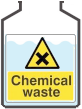
To empty a cuvette, select it and then click on the
 waste container.
To switch the spectrophotometer on and off, click on the
![]() switch
switch
To access the cuvette compartment, click on the  latch or on the cover.
To introduce a cuvette into the spectrophotometer or to get it out:
- Select a cuvette clicking on it or on the radio button below it.
- Click on one of the arrows

- (Beware that the compartment cover must be open)
Action of buttons and icons:
 records the measurement in the external monitor
 records a spectrum
 clears all information off the monitor
  captures an snapshot of the spectrum,
which can then be copied or saved via the context menu in the browser (*),
or captures the list of absorbance measurements in the monitor in a format more suitable for copying.
captures an snapshot of the spectrum,
which can then be copied or saved via the context menu in the browser (*),
or captures the list of absorbance measurements in the monitor in a format more suitable for copying.
-
 represents the waste disposal vessel;
when clicked on it, the selected cuvette will be emptied.
represents the waste disposal vessel;
when clicked on it, the selected cuvette will be emptied.
The pipette delivers 1 mL aliquots. Each cuvette has a 3 mL capacity; if surpassed, it will spill (the cuvette will then be emptied in order to continue). To be measured in the spectrophotometer, the cuvette must contain at least 2 mL of sample.
The absorbance reading displays ##### whenever it is not possible to measure: if the compartment cover is open or if the volume in the cuvette is not enough.
*) Help about saving or pasting the "photograph" of results:
- Usually the web browser offers an option in the context menu to “save image” which will save it to a PNG format file (right-clicking on the image).
- In the same menu there is usually an option to “copy image”. It may then be pasted in another program, but it may not work with the regular “paste”; try “special paste” and choose “bitmap” or “PNG image”.
- All these is not possible on the graph shown in the monitor below the spectrophotometer,
it only works on the "photo" that pops in the centre of the window when you click on the
icon.
Disponible también en español
Plant pigments, experiment 1: absorption spectra of pigments (standards)
Aim: register and compare the spectrum (absorption of visible light) of diverse plant pigments: chlorophyll a, chlorophyll b and β-carotene.
Materials: We will use solutions of three pure pigments (chlorophyll a, chlorophyll b and β-carotene standards) in a suitable solvent.
Procedure:
- Calibrate the spectrophotometer with the solvent: for this, fill a cuvette with 3 mL of the solvent, take it into the spectrophotometer, press the “A=0” button to set zero absorbance and so avoid any interference by the solvent on the absorption spectra of pigments. Take the cuvette out and discard its contents.
- Run the spectral scan for each pigment: to do this, fill each cuvette with 3 mL of one pigment.
Take each cuvette, one at a time, into the spectrophotometer, press the
button
to run the spectral scan and obtain the absorption spectrum. Save each image obtained by
clicking on , in order to
compare the spectra once you finish the experiment and to include the images in you lab report with
the coorresponding caption.
- To analyse each spectrum directly you may move around using the arrows at the side of the wavelength display (▲▼) and check the absorbance value for each λ.
Analysis of results (you must include questions and answers in your lab report)
- Paste the image of each absroption spectrum, quoting to which pigment it correspond.
- Analyse the spectrum for each pigment and write down two wavelengths for which there is an absorption peak (local maximum). Additionally, write down the absorbance for each peak (you may use the following table).
- Observe and compare the molecular structure of chlorophyll a and chlorophyll b
displayed in the "rationale" section (further down in this page).
a. Do you appreciate very different structures? Explain which is the difference between both.
b. Explain whether the difference is manifested in the absorption spectra. - Observe the molecular structure of β-carotene and compare with the chlorophylls.
a. Do you appreciate very different structures? Explain which is the difference between them.
b. Explain whether the difference is manifested in the absorption spectra of the 3 compounds. - Explain if recording the absorption spectrum of a substance could serve to identify or characterise compounds which are different.
Rationale
Radiant energy from the sun is stored in photosynthetic organisms where pigments such as chlorophyll and other associated pigments act as molecular traps for ultraviolet and visible radiant energy.1.
Chlorophyll is the green pigment in plants and plays a vital role in photosynthesis, the process by which plants convert water, carbon dioxide and sunlight (one of the forms of energy) into carbohydrates. Chlorophyll a is predominant in plants, although chlorophyll b may also be present. The two pigments differ slightly in structure1,2, as can be observed in structures in Figure 1.
Carotenoids are another important group of photosynthetic pigments, and can be classified into carotenes and xanthophylls. Carotenoids are generally responsible for the yellow or orange colours of flowers, fruits and roots. The most common carotene is β-carotene1,2.
In an absorption spectrum one may correlate the intensity of absorption due to the pigment with the wavelength of each radiation which has been beamed on the solution. In such spectrum it is common to observe absorption maxima (peaks) at a certain wavelength, which are characteristic of each compound. The variable that measures the intensity of absorption is called absorbance (A)1.
| chlorophyll a | chlorophyll b | β-carotene |
 |
 |
 |
References:
- Díaz de Vivar, E.; Solís, M. and Avaro, M. 2018. Química orgánica: de la arquitectura molecular a la función biológica. Comodoro Rivadavia: EDUPA. ISBN: 978-987-1937-92-9. Available online.
- Mancilla, E.; Castrejón, R., Rosas, M.; Blanco, Z. and Pérez J. 2013. Extracción y separación de pigmentos vegetales. México. Universidad del Valle de México.
Plant pigments, experiment 2: absorption spectra of pigments separated by column chromatography
Overall aim: to identify or characterise, through their absorption spectra, pigments preent in a plant extract and in its 2 fractions separated by chromatography.
Write down in your laboratory notebook the code assigned to your experiment
(in this particular case it is ???).
Specific aims:
- To analyse the absorption spectra of the fractions obtained from plant extracts after subjecting these to adsorption column chromatography.
- To compare the absorption spectra of the plant pigment fractions among them.
- To identify compounds present in each chromatographic fraction, by comparison of their absorption spectra with those of the pigment standards: β-carotene and chlorophyll a.
- To compare the absorption spectra of chromatographic fractions with that of the whole extract before chromatography.
Materials: We will use solutions of two pure pigments (chlorophyll a and β-carotene standards) and two fractions with plant pigments obtained after separation by column chromatography from spinach, chard or microalgae extracts.
Design of the experiment:
- Calibrate the spectrophotometer with the solvent: for this, fill a cuvette with 3 mL of the organic solvent as reagent blank, take it into the spectrophotometer, press the “A=0” button to set zero absorbance. Take the cuvette out and discard its contents.
- Run the spectral scan: to do this, fill each cuvette with 3 mL of one of the samples.
Take each cuvette, one at a time, into the spectrophotometer and press the
button
to run the spectral scan and obtain the absorption spectrum. Save each image obtained by clicking on
,
in order to compare the spectra once you finish the experiment and to include the images in you lab report
with the coorresponding caption.
- To analyse each spectrum directly you may move around using the arrows at the side of the wavelength display (▲▼) and check the absorbance value for each λ.
Analysis of results:
- Analyse the absorption spectra obtained for each sample quoted in the following table
(which you should record in your notebook and lab report), stating the wavelength of the local maxima (λ)
within the range 350 to 750 nm.
Table 2. Data record for the analysis of absorption spectra of the pigment standards and extracts from plant samples. cuvette nr. Wavelength of
local maxima (λ, nm)β-carotene standard chlorophyll a standard Plant extract 1st chromatographic fraction 2nd chromatographic fraction - Considering the absorption spectra of the standards (chlorophyll a and β-carotene):
a. What can you say about the absorption spectrum of the first chromatographic fraction?
b. What can you say about the absorption spectrum of the second chromatographic fraction?
c. What can you say about the absorption spectrum of the plant whole extract?
d. If the first fraction was eluted from the chromatographic column using hexane as solvent and the second one using acetone, what can you say about the polarity of the different pigments? Suggestion: analyse the structure of the standards in figure 1 of the first sub-tab in the pigments tab.
Plant pigments, experiment 3: relation between absorbance and concentration
Aim: To analyse the effect of concentration of a plant pigment on it absorption through the analysis of its spectrum.
Calibration: for this, fill a cuvette with 3 mL of the organic solvent as reagent blank, take it into the spectrophotometer and press the “A=0” button to set zero absorbance. Take the cuvette out and discard its contents.
Design of the experiment:
- Using one of the pigments and the solvent, prepare 3 samples in the respective cuvvetes, so that they contain
different volumes of the pigment, always with the same final volumen of 3 mL.
You may organise yourself by filling in the following table with the volumes to be placed in each cuvette:
Table 3A. Record of preparation of samples to analyse the relation between absorbance and concentration of the pigment Pigmento selected: Cuvette number: Volume of pigment (mL): Volume of solvent (mL): - Once the 3 cuvettes have been prepared, take a photo to be included in your report and, also, include both in your notebook and in the lab report the values you have entered in the preceding table.
- Scan the spectrum for each of the solutions: for this, take each cuvette, one at a time, into the
spectrophotometer and press the button
to record the absorption spectrum of each dilution. Save the image obtained clicking on
, so you can compare the spectra
when you finish the experiment, as well as include them in your report.
Analysis of results (include these questions and answers in your lab report)
- Analyse the absorption spectra obtained for each solution and compare them
against each other:
Measure and write down the intensity of absorbance and the wavelength of the two highest peaks for each sample, following this table:Table 3B. Analysis of the absorption spectra First peak Second peak cuvette number Wavelength (λ1, nm) Absorbance Wavelength (λ2, nm) Absorbance - Describe the effect of diluting the selected pigment.
- Does the position of λ of the maximum absorption peaks change? Explain your answer.
Plant pigments, experiment 4: quantitation of pigment concentration in an unknown sample.
Aim: to prepare a calibration curve with one of the pigments in order to measure the unknown concentration in a sample of interest.
Materials: A solution of chlorophyll a will be used. Later you can repeat the assay with othe rpigments, but choosing the right wavelength for their quantitaion: λ= 663 nm for chlorophyll a and λ= 480 nm for β-carotene.
Calibration: First, fill a cuvette with 3 mL of solvetn (90% acetone in water), take the cuvette
into the spectrophotometer, adjust the suitable wavelength and press the “A=0” button. Take the cuvette out and
discard its contents.
If you change the wavelength to measure another pigment, you must repeat the calibration.
To make dilutions with adequate precision, in this esperiment a double flush bulb pipette will be used rather than a
Pasteur pipette:

~~ 1st part: chlorophyll a ~~
Design of the experiment: Using chlorophyll a and solvent, prepare 3 samples in the 3 cuvettes, containing different volumes of chlorophyll a and always the same total volume of 3 mL.
To help you do this, fill previously this table in (or better, ona you make in your lab notebook) with the volumes you intend to add into each cuvette:
| cuvette: | 1 | 2 | 3 | |
| volume of chlorophyll a: | mL | |||
| volume of solvent: | mL |
Once the 3 mixtures are ready in the 3 cuvettes, take these one at a time into the spectrophotometer
and write down the absorbance measurements. You may also press the
button to record each absorbance value in the
monitor under the spectrophotomoeter.
| cuvette: | 1 | 2 | 3 |
| A663 = |
Qualitative analysis of results: Observe the relation between colour in the cuvettes and the absorbance value. Comparing the 3 measurmente, do you perceive some dependency between the value of absorbance and the higher or lower concentration of chlorophyll in the cuvette?
Quantitative analysis of results: Calculate the concentration of chlorophyll a in the final mixture in each cuvette, knowing the concentration if the bottle is 1 mg/L. Prepare a table with the data and make a graph plotting A (absorbance) against C (concentration. How would you describe your observations?
| cuvette | C (mg/L) |
A663 | A663
Cchlorophyll a (mg/L)
|
| 1 | |||
| 2 | |||
| 3 |
Using the graph, solve this problem:
2 mL of a problem sample are mixed with 1 mL of solvent. Calculate the concentration chlorophyll a in the
original sample if the assay has yielded a value
A663 = disclose the value
Enter your result: C =
mg/L and check if it is correct.
~~ 2nd part: β-carotene ~~
Repeat the whole process with β-carotene, measuring in this case absorbance at 480 nm (concentration of carotene in the bottle is 1 mg/L).
(Calibration + design of the experiment + analysis of results)
Using the graph, solve this problem:
1 mL of a problem sample are mixed with 2 mL of solvent. Calculate the concentration of β-carotene in the
original sample if the assay has yielded a value
A480 = disclose the value
Enter your result: C =
mg/L and check if it is correct.
~~ 3rd part: unknown sample ~~
Write down in your lab notebook the code assigned to your experiment (which in this particular case it is
???).
Let us analyse both the whole plant extract and its two fractions obtained with chromatography (please read the Pigments 2: extracts tab for more information)
Take into account that, if the absorbance obtained in any assay was higher than the maximum one obtained in the calibration line for the same wavelength, you should not use such datum. It will be necessary to prepare in an empty cuvette a sample more diluted than what was indicated, so that its absorbance falls within the range of the calibration line. Obviously, such new dilution must be taken into account for the subsequent calculation of concentration.
a) Plant extract
Mix into the cuvette 2 mL of the extract with 1 mL of solvent. Measure its absorbance at both 480 and 663 nm (before measuring at each wavelength you need calibrate A=0 for that same wavelength, using another cuvette filled with just solvent).
Calculate the concentrations of both chlorophyll a and β-carotene in the extract:
mg/L of chlorophyll a, and
mg/L of β-carotene
Check if it is correct.
b) Fraction 1
Mix into a cuvette 2 mL of fraction 1 with 1 mL of solvent. Measure the absorbance at both 480 and 663 nm (before measuring at each wavelength you must calibrate A=0 for that same wavelength, using another cuvette filled with just solvent).
Calculate the concentrations of chlorophyll a and β-carotene in fraction 1:
mg/L of chlorophyll a, and
mg/L of β-carotene
Check if it is correct.
b) Fraction 2
Mix into a cuvette 2 mL of fraction 2 with 1 mL of solvent. Measure the absorbance at both 480 and 663 nm (before measuring at each wavelength you must calibrate A=0 for that same wavelength, using another cuvette filled with just solvent).
Calculate the concentrations of chlorophyll a and β-carotene in fraction 2:
mg/L of chlorophyll a, and
mg/L of β-carotene
Check if it is correct.
Disponible también en español